Why Ultrasound?
We are sorry, but we cannot make appointments directly with pet owners. Appointments need to be scheduled by your veterinarian. Please contact your regular veterinarian to request that they contact Mettasound for an ultrasound examination. If you are looking for a new family vet or need ultrasound services, just contact us for recommendations!
People are most familiar with the type of ultrasonography known as a sonogram which allows a sonographer or physician to view the fetus of a pregnant woman. Many people have also heard of an echocardiogram which is an ultrasound of the heart.
Ultrasound is like ordinary sound except it has a frequency higher than humans can hear. The sound is reflected off of internal structures. The returning echoes are then received by the transducer and converted by an electronic instrument into an image on a monitor. It is similar to how the sonar on a ship works. Extensive training is required in order to correctly obtain and interpret these images.
An ultrasound is exam is not painful, however, slight discomfort from pressure may be experienced. In general, animals do not need to be anesthetized for this procedure, but sedatives may be necessary for those animals that are aggressive or anxious. A water soluble gel and sometimes alcohol is applied over the area to be examined and the transducer is placed on the skin (the pet’s hair coat over the area to be examined will typically need to be shaved). Ultrasonography may also be used in guiding the needle for sampling of internal organs and centesis.
Widespread use in human and veterinary medicine for many years has not revealed any harmful effects with the use of ultrasonography.
What can be seen with ultrasound?
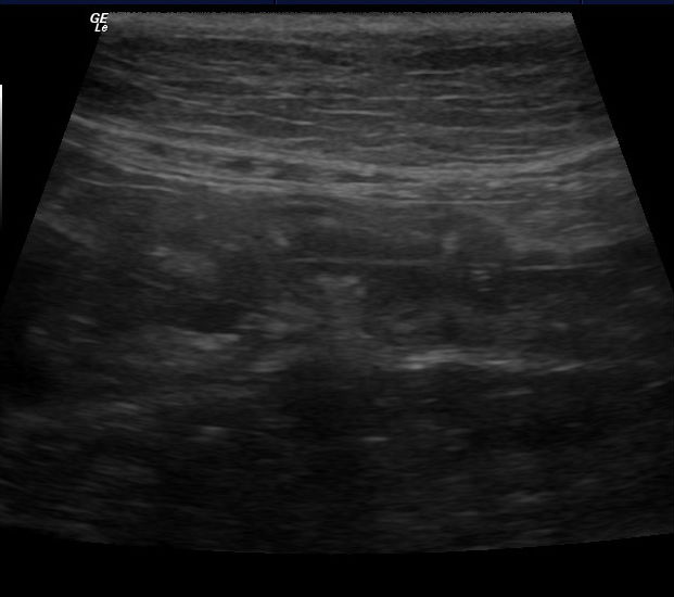
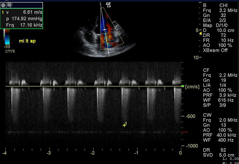
Ultrasound is used to diagnose a variety of both benign and malignant diseases such as the presence of bladder stones gall bladder, prostate or kidneys, the presence of enlarged lymph nodes, abnormal blood vessels, or free fluid within the abdomen. It is especially good in diagnosing disease of the pancreas (pancreatitis), adrenal abnormalities, urinary bladder wall tumors, and uterine infections (pyometra). With ultrasound guided sample acquisition, ultrasound can often differentiate benign organ changes and cancer. In animals with a history of vomiting, ultrasound can be used to evaluate if the problem is within the liver, gallbladder or pancreas. It can often diagnose problems that are associated with the stomach or small intestinal wall, or see an intestinal foreign body, thus preventing a labor intensive and costly upper GI barium study. In the heart (echocardiography) ultrasound is at it’s best, as the heart is a fluid filled organ. Abnormalities such as a diseased heart muscle (hypertrophic and dilatory cardiomyopathy), fluid around the heart (pericardial effusion), valvular disease and congenital abnormalities can be diagnosed and the severity assessed. Often, your vet will suggest an echocardiogram if a heart murmur is heard. Visualizing the heart with ultrasound will pinpoint where the murmur is coming from and if there is a need for medication. It also allows for a baseline to follow progression of disease. Heartbase tumors, which are rarely seen on radiographs, are also easily visualized with ultrasonography.
Ultrasound is often used in conjunction to diagnose complex conditions and as such, it’s best to think of ultrasound as a piece of the puzzle and not the only diagnostic test your pet may need. Ultrasound doesn’t replace radiographs, nor do radiographs replace ultrasound. They both complement each other by showing different things. For example, ultrasound is not good at assessing the lungs, but radiographs are. A radiograph may show an enlarged heart, but an echocardiogram will be able to tell your vet why the heart is enlarged. Again, it’s all pieces to the puzzle of a diagnosis. Many times an ultrasound will be normal and not yield a specific explanation for the clinical signs your pet may be exhibiting, but it helps your veterinarian rule-out significant differentials which is important so that the time to a diagnosis isn’t wasted chasing down the wrong answers.
There are no confirmed biological effects on patients or instrument operators caused by exposures from present diagnostic ultrasound instruments. Although the possibility exists that such biological effects may be identified in the future, current data indicate that the benefits to patients of the prudent use of diagnostic ultrasound outweigh the risks, if any, that may be present.